


قیمت 19,000 تومان
عملکرد الکترومایوگرافی عضلات شانه
ادبیات و پیشینه تحقیق عملکرد الکترومایوگرافی عضلات شانه در وضعیت
الکترومایوگرافی عضلات شانه
فصل دوم: پیشینه پژوهش عضلات شانه
2-1- مقدمه عضلات شانه :
2-2-مبانی نظری ..
2-2-1-ساختار استخوانهای تشکیل دهنده کمربندشانه ای..
2-2-1-ساختار استخوانهای تشکیل دهندهی کمربند عضلات شانه ..
2-2-1-1 استخوان بازو:
2-2-1-2- کتف:
2-2-1-3-ترقوه:
2-2-2-ساختار مفاصل و بافتهای حمایت کننده کمربند شانهای..
2-2-2-1-جناغی-ترقوهای..
2-2-2-2-آخرومی-ترقوهای..
2-2-2-3-کتفی-صدری..
2-2-2-4-مفصل تحت دلتوئید.
2-2-2-5-مفصل گلنوهومرال.
2-2-3-پایدارکنندههای استاتیکی مفصل گلنوهورمرال:
2-2-3-1- سطح مفصلی..
2-2-3-2- لابرم:
2-2-3-3- کپسول مفصلی:
2-2-3-4- لیگامنتها:
2-2-4- پایدارکنندههای دینامیکی مفصل گلنوهومرال.
2-2-4-1- عضلات RC:
2-2-4-2-زوج نیرو صفحه فرونتال.
2-2-4-3-زوج نیروی صفحه عرضی..
2-2-5-حرکات و دامنه حرکتی شانه.
2-2-5-1-فلکشن و آبداکشن..
2-2-5-2-اداکشن و اکستنشن..
2-2-5-3-چرخش داخلی و خارجی..
2-2-5-4-فلکشن افقی و اکستنشن افقی..
2-2-6-حرکات ترکیبی کمربند شانه.
2-2-7-فعالیت عضلانی در کمربند شانه.
2-2-7-1-عضلات اسکاپولاهومرال.
عضله دلتوئید.
عضلات روتاتورکاف…
عضله فوقخاری:
عضله تحتخاری..
عضله گردکوچک….
عضله تحتکتفی..
2-2-7-2-عضلات آکسیو اسکاپولار و آکسیوکلاویکولار.
عضله ذوزنقه:
بالابرنده کتف:
عضله دندانهای قدامی..
سینهای کوچک:
2-2-7-3-عضلات آگسیوهومرال.
عضله سینهای بزرگ…
2-3- پیشینه پژوهش:
2-3-1-آسیب عضلات روتاتورکاف:
2-3-2-ناپایداری مفصل گلنوهومرال.
2-3-3-دسته بندی سندرومهای برخوردی..
2-3-4- تستهای سندرومهای برخوردی:
2-3-3-1-تست برخوردی نیر.
2-3-3-2-هاوکینز.
2-3-3-4-تست جاب…
2-3-3-4-تست سوپینیشن..
2-3-5-مروری بر تحقیقات گذشته .
منابع عضلات شانه
الکترومایوگرافی عضلات شانه
ساختار استخوانهای تشکیل دهنده کمربند عضلات شانه
کمپلکس عضلات شانه از استخوانهای ترقوه[1]، کتف[2] و بازو[3] تشکیل شده است و به طور پیچیده ای ترکیبی از سه مفصل را ایجاد میکنند(اتیس2004[4]).(شکل2-1)
ساختار استخوانی مفصل گلنوهومرال شامل یک سطح مفصلی بازویی-دوری کوچک، که با سر بزرگ استخوان بازو مفصل میشود به تثبیت کنندههای لیگامنتی و عضلانی آن در سراسر قوس حرکتی نیاز دارد. آسیب یا استفاده بیش ازحد، پایدار کنندههای استاتیکی و دینامیکی باعث میشود که شانه بیشتر در معرض خطر آسیب باشد. 8 تا 20% آسیبهای ورزشی آسیب شانه است(استین برک[5] 1999; پائول جی دبیلیو و بابرفاس[6] 1999)
در اینجا کمربند عضلات شانه از نقطه نظر ساختاری به شرح زیر برسی میشود:
- آناتومی استخوانی (بازو، کتف، ترقوه)
- مفاصل (بازویی-دوری، کتفی-صدری، آخرومی-ترقوه ای و جناقی-چنبری)
- پایدارکنندههای استاتیکی (سطح مفصلی،لابرم، کپسول و لیگامنت)
- عضلات پایدار کنندههای دینامیکی (روتاتورکاف، دلتوئید و پایدار کنندههای کتف)
اگرچه این قسمتها را به صورت مجزا بررسی میکنیم اما آنها برای تولید حرکت عضلات شانه به عنوان یک واحد دینامیک، متکی به هم عمل میکنند.
شناخت آناتومیکی عملکردی شانه به همراه بررسی منابع مکرر آسیب شانه به متخصصین این اجازه را میدهد که رویکردی را برای مراقبت از شانه بدست بیاورند(تری و چاپ 2000[7]).
[1]-clavicle
[2]-Scapulae
[3]-Humerus
[4] Oatis
[5] steinbarek
[6] Powell JW and barber foss
[7] Terry and Chopp
جهت مشاهده و دانلود مطالب درباره الکترومایوگرافی کلیک کنید .
ترقوه – عضلات شانه
استخوان ترقوه از طریق مفصل جناغی-چنبری تنه را به کمربند شانه متصل میکند. ترقوه در امتداد محور طولی خود 2 تا قوس دارد. 3/1 بیرونی آن پهن و محلی برای اتصال عضلات و لیگامنتها است در حالی که 3/1 داخلی لولهای است که بار محوری را تحمل میکند (شکل 2-7).
| شکل2-7- استخوان ترقوه |
ناحیه 3/1 میانی به طور مکانیکی ضعیف و بخش باریکی است که اغلب شکستگیها در این ناحیه اتفاق میافتد. ترقوه به عنوان محل نشست عضلات، محافظ اعصاب و رگهای خونی است و دامنه حرکت شانه را افزایش میدهد (لجانگرن1972[1]). بعلاوه ترقوه از انتقال تحتانی کمربند شانه، توسط لیگامنت غرابی-ترقوهای جلوگیری میکند(راکود 1991[2]).
ساختار مفاصل و بافتهای حمایت کننده کمربند شانهای
کمربند شانه تعداد زیادی مفصل دارد که هرکدام در حرکت بازو نقش دارند. حرکت بازو در مفصل گلنوهومرال رخ میدهد در حالی که ممکن است مقدار کمی حرکت در هریک از مفاصل دیگر نیز رخ دهد(رحمانزاده 1380)مفاصل کمربند شانه شامل:
جناغی-ترقوهای[3]
آخرومی-ترقوهای[4]
کتف با قفسه سینه[5]
مفصل تحت دلتوئید[6]
گلنوهومرال[7]
همه مفاصل جز مفصل کتف با قفسه سینه و مفصل تحت دلتوئید(بین اجزای موجود در زیر عضله دلتوئید)، مفاصل سینوویال هستند. در مفصل کتف با قفسه سینه ومفصل تحت دلتوئید به خاطر اینکه این دو ساختار مفصلی مستقیما با هم تماس ندارند. بین کتف و قفسه سینه بیشتر عضلات قرار گرفتهاند تا غضروف و بافت لیفی بنابراین این مفصل جزء دسته بندی مفاصل به حساب نمیآید ولی به دلیل حرکات مکرر بین کتف و قفسه سینه میتوان واژه مفصل فیزیولوژیکی را به آن نسبت داد. مفاصل فیزیولوژیکی درواقع عهدهدار ارتباط فیزیولوژیکی بین اجزای مختلف هستند(رواسی 1386; اتیس[1]2009).
[1] Otis
[1] Ljunggren
[2] Rockwood
[3] Sternoclavicular
[4] Acromioclavicular
[5] Scapulothoracic
[6] Subdeltoid
[7] Glenohumeral
پایدارکننده های دینامیکی مفصل گلنوهومرال
پایدارکنندههای دینامیکی، سطح مفصلی، لابرم و فشار درون مفصلی[1] در میانه حرکت نقش پایداری مفصل را برعهده دارند درحالیکه لیگامنتهای مفصل گلنوهومرال در میانه دامنه حرکتی شل هستند (آبود و ساسلواسکی2002[2]).
اگرچه پایدارکنندههای دینامیکی در میانه حرکت مهم هستند اهمیت آنها بیشتر در حفظ ثبات مفصل در انتهای دامنه حرکتی است(لیبریل و لی 2005[3]).
[1] intra-articular pressure
[2] Abboud and Soslowsky
[3] Labriola, Lee
حرکات ترکیبی کمربند شانه
همانطور که قبلا بیان شد تمام مفاصل شانه باید با همدیگر کار کنند تا حرکت بازو را فراهم کنند. در 30 درجه اول آبداکشن یا 45-60 درجه فلکشن کتف از ستون مهرهها دور میشود تا وضعیت ثابتی را روی قفسه سینه پیدا کند بعد از ثبات، کتف چرخش فوقانی، آبداکشن و الویشن پیدا میکند و ترقوه به صورت خلفی بالارفته و پرتراکشن میدهد تا بازو بتواند آبداکشن و فلکشن را انجام دهد(اینهورن 1985[1]).
در مراحل اول آبداکشن و فلکشن حرکات در مفصل گلنوهومرال انجام میشوند بعد از 30 درجه آبداکشن یا 60-45 درجه فلکشن نسبت حرکت بین کتف و بازو 5:4 است یعنی به ازای 5 درجه حرکت در مفصل گلنوهومرال 4 درجه کتف میچرخد و در کل دامنه حرکتی این نسبت تقریبا 2:1 است(هلبچ 1985). در 180 درجه آبداکشن، 120 درجه در مفصل گلنوهومرال انجام میشود، در کتف 20 درجه در مفصل آخرومی-ترقوهای و 40 درجه در مفصل چناغی چنبری انجام میشود.
هنگامی که بازو به آبداکشن 90 درجه دارد برجستگی بزرگ استخوان بازو به قوس آخرومی ترقوهای برخورد میکند که که بافت نرم زیرین را فشار میدهد. اگر بازو به صورت 30 درجه چرخش خارجی پیدا کند برجستگی بزرگ از زیر قوس آخرومی رها میشود(اینهورن 1985).
اگر بازو به آرامی پایین آورده شود، حرکات آداکشن یا اکستنشن بازو همراه ریتراکشن، دپرشن و چرخش پایینی کتف است.
فعالیت عضلانی در کمربند شانه
عضلات کمربند شانه برحسب نقطه اتصال و مفاصلی که اثر میکنند به سه دسته تقسیم شدهاند:
- عضلات اسکپولا هومرال
2- عضلات آگسیوهومرال
- عضلات آکسیواسکپولار وآکسیوکلاویکولار
جهت مشاهده نمونه های دیگر از فصل دوم پایان نامه تربیت بدنی کلیک کنید.
.
- نمونه ای از فهرست منابع الکترومایوگرافی عضلات شانه
- رواسی، علی اصغر. حرکت شناسی (فیزیولوژی مفاصل) انتشارات نرسی 1386
- طرح سنجش ساختار اسکلتی بدن دانشآموزان دختر مقطع راهنمایی شهر تهران، کارشناس تربیت بدنی دختران شهر تهران حمانزاده، منیژه سادات(1380)
- کارلارد، هینکل. مبانی آناتومی و حرکت. ترجمه دبیدی روشن، ولی الله.انتشارات سمت.تهران1381
- Abboud, J. A. and L. J. Soslowsky (2002). “Interplay of the static and dynamic restraints in glenohumeral instability.” Clinical orthopaedics and related research 400: 48-57.
- Al Levangie PK, Norkin CC (2005). Upper extremity joint complex. Joint structure and function: Comperhensive analysis, philadelphia:F.A.David Company.
- Badarudeen, S. and S. Sabharwal (2008). “Readability of patient education materials from the American Academy of Orthopaedic Surgeons and Pediatric Orthopaedic Society of North America web sites.” The Journal of Bone & Joint Surgery 90(1): 199-204.
- Bigliani, L. (1986). “The morphology of the acromion and its relationship to rotator cuff tears.” Orthop. Trans. 10: 228.
- Bigliani LU, C. E., Butters KP (1991). “Fractures of the shoulder, part I: fractures of the proximal humerus. In: Rockwood CA Jr, Green DP,
- Bucholz RW, eds. Rockwood and Green’s Fractures in Adults.” ed. Philadelphia, PA: JB Lippincott 1. 3rd871-927.
- Bigliani LU, C. E., Butters KP (1991). “Fractures of the shoulder, part I:fractures of the proximal humerus. In: Rockwood CA Jr, Green DP,
- Bucholz RW, eds. Rockwood and Green’s Fractures in Adults.” ed. Philadelphia, PA: JB Lippincott 1: 871-927.
- Blakely, R. L. and M. L. Palmer (1984). “Analysis of rotation accompanying shoulder flexion.” Physical Therapy 64(8): 1214-1216.
- Boardman III, N. D., R. E. Debski, et al. (1996). “Tensile properties of the superior glenohumeral and coracohumeral ligaments.” Journal of Shoulder and Elbow Surgery 5(4): 249-254.
- Boettcher, C. E., K. A. Ginn, et al. (2008). “Standard maximum isometric voluntary contraction tests for normalizing shoulder muscle EMG.” Journal of orthopaedic research 26(12): 1591-1597.
- Boettcher, C. E., K. A. Ginn, et al. (2009). “The ‘empty can’and ‘full can’tests do not selectively activate supraspinatus.” Journal of Science and Medicine in Sport 12(4): 435-439.
- Broome, H. and J. Basmajian (1971). “The function of the teres major muscle: an electromyographic study.” The Anatomical Record 170(3): 309-310.
- Brukner, P. K. K. (2007). clinical sports medicine, McGraw-Hill.
- Cavallo, R. J. and K. P. Speer (1998). “Shoulder instability and impingement in throwing athletes.” Medicine and science in sports and exercise 30(4 Suppl): S18.
- Cole, B. and J. Warner (1999). “Anatomy, biomechanics, and pathophysiology of glenohumeral instability.” Disorders of the shoulder. Diagnosis and management. Philadelphia Baltimore: 208-219.
- Cooper, D. E., S. Arnoczky, et al. (1992). “Anatomy, histology, and vascularity of the glenoid labrum. An anatomical study.” The Journal of bone and joint surgery. American volume 74(1): 46.
- Craig E. Boettchera , K. A. G. (2008). “The ‘empty can’ and ‘full can’ tests do not selectively activate supraspinatus.” Journal of Science and Medicine in Sport 12 ;435:439.
- Adkinson DP., A. (1998). Shoulder Biomechanics. In: Steinbach LS, Petrfy CG, Tir- man PFJ, Feller JF, Gardner GM, editors. Shoulder magnetic resonance imaging., Philadelphia: Lippincott-Raven Publishers.
- Einhorn, A. (1985). “Shoulder rehabilitation: equipment–modifications.” The Journal of orthopaedic and sports physical therapy 6(4): 247.
- EL, Flatow (1988). “Shoulder anatomy and biomechanics. In: Post M, Flatow EL, Bigliani LU, Pollock RG. The Shoulder: Operative Technique. Baltimore, MD: Williams & Wilkins.” (1-42).
- Ellenbecker, T. S. (2004). Clinical examination of the shoulder, WB Saunders.
- ELLMAN, H. (1990). “Diagnosis and treatment of incomplete rotator cuff tears.” Clinical orthopaedics and related research 254: 64-74.
- Favre, P., H. A. C. Jacob, et al. (2009). “Changes in shoulder muscle function with humeral position: A graphical description.” Journal of Shoulder and Elbow Surgery 18(1): 114-121.
- Funk, L. (2005). “Rotator Cuff Biomechanics. For MSc Orthopaedic Engineering.”
- Gerber, C., O. Hersche, et al. (1996). “Isolated Rupture of the Subscapularis Tendon. Results of Operative Repair*.” The Journal of Bone & Joint Surgery 78(7): 1015-1023.
- Gerber, C. and R. J. Krushell (1991). “Isolated rupture of the tendon of the subscapularis muscle. Clinical features in 16 cases.” Journal of Bone & Joint Surgery, British Volume 73(3): 389-394.
- Gibb, T. D., J. A. Sidles, et al. (1991). “The effect of capsular venting on glenohumeral laxity.” Clinical orthopaedics and related research(268): 120.
- Gschwend, N., D. Ivošević-Radovanović, et al. (1987). “Rotator cuff tear—relationship between clinical and anatomopathological findings.” Archives of Orthopaedic and Trauma Surgery 107(1): 7-15.
- Halbach, J. W., Tank, R. T (1985). The shoulder. In J. A. Gould, G. J. Davies (Eds.), Orthopaedic and Sports Physical Therapy. St. Louis: C.V. Mosby,.
- Halder, A. M., E. Itoi, et al. (2000). “Anatomy and biomechanics of the shoulder.” The Orthopedic clinics of North America 31(2): 159.
- Hall, M. C. and M. Rosser (1963). “The structure of the upper end of the humerus with reference to osteoporotic changes in senescence leading to fractures.” Canadian Medical Association Journal 88(6): 290.
- Hawkins, R. and P. Hobeika (1983). “Impingement syndrome in the athletic shoulder.” Clinics in sports medicine 2(2): 391.
- Hawkins, R. and J. Kennedy (1980). “Impingement syndrome in athletes.” The American journal of sports medicine 8(3): 151-158.
- Hulstyn, M. J. and P. D. Fadale (1997). “Shoulder injuries in the athlete.” Clinics in sports medicine 16(4): 663-679.
- Inman, V. T. and L. R. C. Abbott (1944). “Observations on the function of the shoulder joint.” The Journal of Bone and Joint Surgery (American) 26(1): 1-30.
- Itoi E, K. T., Sano A, Urayama M, Sato K. (1999). “Which is more useful, the “full can test”or the“empty can test” indetecting the torn supraspinatus
- tendon?” Am J Sports Med: 65–68.
- Jobe, C. (1998). Gross anatomy of the shoulder In: Rockwood CA Jr, Matsen FA II, eds. The Shoulder., Philadelphia, PA: WB Saunders.
- Jobe, F. W. and C. M. Jobe (1983). “Painful athletic injuries of the shoulder ” Clinical orthopaedics and related research(173): 117.
- Jobe, F. W. and D. R. Moynes (1982). “Delineation of diagnostic criteria and a rehabilitation program for rotator cuff injuries.” The American journal of sports medicine 10(6): 336-339.
- Joe de Beer, K. v., Deepak N Bhatia ((2000 )). “subscapularis tendon tear.” Orthopedic Shoulder Surgeon: 16(17):E18.
- Kamkar, A., J. Irrgang, et al. (1993). “Nonoperative management of secondary shoulder impingement syndrome.” Journal of Orthopaedic and Sports Physical Therapy 17: 212-212.
- Kelly, B. T., W. R. Kadrmas, et al. (1996). “The Manual Muscle Examination for Rotator Cuff Strength An Electromyographic Investigation.” The American journal of sports medicine 24(5): 581-588.
- Konrad, P. (2005). “The abc of emg.” A Practical Introduction to Kinesiological Electromyography 1.
- Kuechle, D. K., S. R. Newman, et al. (2000). “The relevance of the moment arm of shoulder muscles with respect to axial rotation of the glenohumeral joint in four positions.” Clinical Biomechanics (Bristol, Avon) 15(5): 322-329.
- Kronberg, M., G. Nemeth, et al. (1990). “Muscle activity and coordination in the normal shoulder. An electromyographic study.” Clin Orthop 257: 76-85.
- Kvitne, R. S. and F. W. Jobe (1993). “The diagnosis and treatment of anterior instability in the throwing athlete.” Clinical orthopaedics and related research(291): 107.
- Labriola, J. E., T. Q. Lee, et al. (2005). “Stability and instability of the glenohumeral joint: the role of shoulder muscles.” Journal of Shoulder and Elbow Surgery 14(1): S32-S38.
- leavangie Pamela K, N. C. C. (2005). “Joint structure and function: a comprehensive analysis. .” F. A. Davis Company. Philadelphia.
- Lippitt, S. B. n., J.E. Harris, S.L. Sidles, J.A. Harryman, D.T. Matsen, F.A. (1993). “Glenohumeral stability from concavity-compression: a quantitative analysis.” Journal of Shoulder and Elbow Surgery 2(1): 27-35.
- Liu, J., R. Hughes, et al. (1997). “Roles of deltoid and rotator cuff muscles in shoulder elevation.” Clinical Biomechanics 12(1): 32-38.
- Ljunggren, A. E. (1979). “Clavicular function.” Acta Orthopaedica 50(3): 261-268.
- Malanga, G. A., Y. Jenp, et al. (1996). “EMG analysis of shoulder positioning in testing and strengthening the supraspinatus.” Medicine and science in sports and exercise 28: 661-664.
- Marks PH, W. J. (1994). “Rotator cuff disorders of shoulder.” J hand Ther: 7;90:98.
- Moseley, H. and B. Overgaard (1962). “The anterior capsular mechanism in recurrent anterior dislocation of the shoulder.” J Bone Joint Surg Br 44: 913-927.
- NEER, C. S. (1983). “Impingement lesions.” Clinical orthopaedics and related research 173: 70-77.
الکترومایوگرافی عضلات شانه
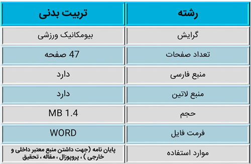
| رشته | تربیت بدنی |
| گرایش | بيومكانيك ورزشی |
| تعداد صفحات | 47 صفحه |
| منبع فارسی | دارد |
| منبع لاتین | دارد |
| حجم | 1.4 MB |
| فرمت فایل | ورد (Word) |
| موارد استفاده | پایان نامه (جهت داشتن منبع معتبر داخلی و خارجی ) ، پروپوزال ، مقاله ، تحقیق |
نقد و بررسیها
هنوز بررسیای ثبت نشده است.
موارد مرتبط


نقد و بررسیها
هنوز بررسیای ثبت نشده است.